

Advanced Third Molar Extraction with Clinical Safety and Predictability
AN INTENSIVE TRAINING PROGRAMME DESIGNED FOR DENTAL PROFESSIONALS WHO WANT TO PLAN AND PERFORM THIRD MOLAR EXTRACCTIONS WITH TECHNICAL CONTROL, CONFIDENCE AND A REAL REDUCTION IN COMPLICATIONS.
30 and 31 May 2026 · 16 hours · Language: Portuguese
Before the scalpel, there is a critical decision
Most complications in third molar surgery do not occur during the procedure.
They occur beforehand — during planning.
Nerve injuries, prolonged trismus, haemorrhage or oroantral communication are rarely caused by a lack of manual skill.
They result from incomplete assessment, imprecise imaging interpretation and poorly structured clinical decisions.
👉 An uncomfortable but essential question:
Can you reliably predict the real surgical difficulty of a third molar before you begin?
When planning fails, risk becomes real
In daily clinical practice, third molar extraction remains one of the procedures with the highest rate of preventable complications.
Proximity to the inferior alveolar nerve
Underestimated difficulty
Inadequate flap design
Excessive osteotomy
Lack of an alternative surgical strategy
Surgical safety is not talent. It is method.
Surgical safety is not talent. It is method.
This course was designed to change how dental professionals think, plan and execute third molar extractions.
The focus is not on “doing more surgeries”, but on doing them better, through:
Structured clinical reasoning
Clear operative sequencing
Evidence-based decision-making
Predictable outcomes in real practice
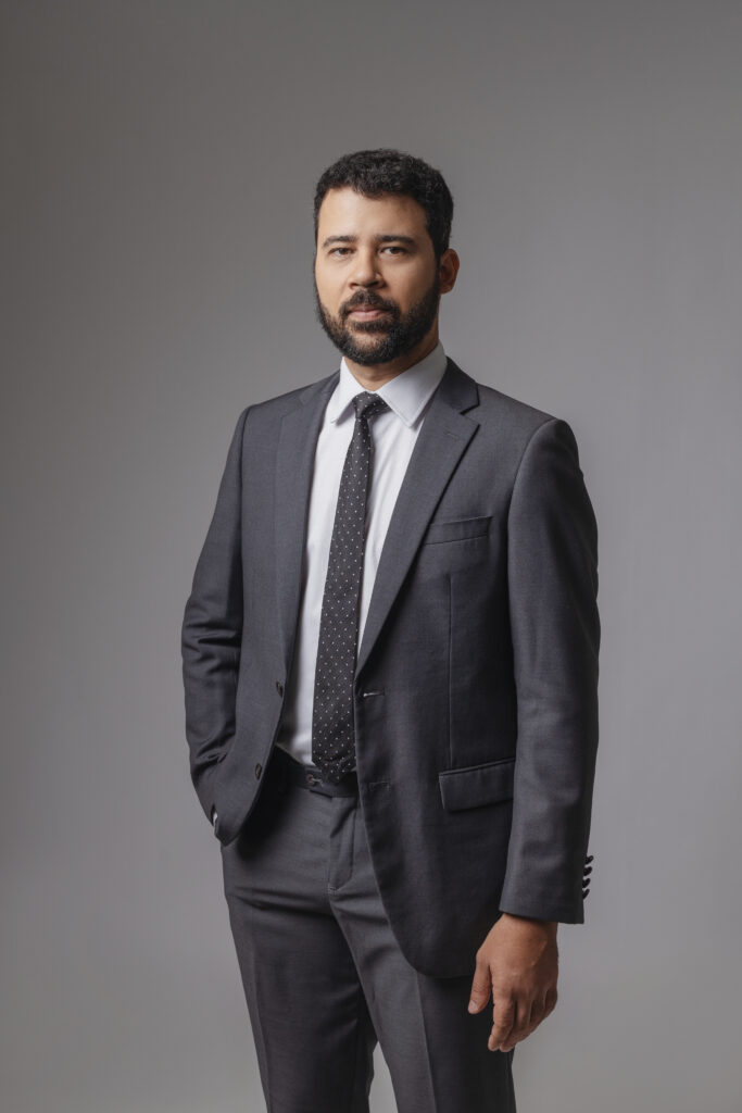
Oral and Maxillofacial Surgeon, PhD in Oral and Maxillofacial Surgery, with strong academic and clinical activity and international recognition.
His teaching approach is known for clarity, rigour and immediate clinical applicability — preparing professionals to act safely in real surgical scenarios.
INVESTMENT IN THIRD MOLAR EXTRACTION
- Face-to-face training
- Small groups
- Close supervision
- Official SIGO certification (EU recognised)
LANGUAGE
Portuguese
DURATION
16 hours
CERTIFICATE
SIGO
CITY
Lisbon
LOCAL
Dental Courses Europe
Advanced surgical planning
Learn how to correctly analyse applied anatomy, impaction classifications and imaging (panoramic X-ray and CBCT), identifying risk factors before surgery.
Structured technical execution
Each surgical stage is addressed step by step: incision → flap design → osteotomy → odontosection → extraction → suturing.
Prevention and management of complications
Understand how to prevent — and how to manage — the most common complications associated with third molar surgery.
Supervised hands-on training
Intensive practical training in a controlled environment, with continuous guidance and correction.
Live surgical demonstration
Observe the real application of the method in a clinical setting, from planning to post-operative management.
Accurate case selection
Advanced interpretation of panoramic X-rays and CBCT
Predictable surgical planning
Atraumatic extraction techniques
Reduction of surgical risk
• Greater clinical confidence in daily practice
- Dental professionals who perform or wish to perform third molar extractions
- Clinicians seeking greater surgical safety and predictability
- Professionals aiming to reduce complications
- Those looking for theory-only training
- Those unwilling to apply the techniques in clinical practice
Applied Intensive Theory
Anatomy, classifications, surgical planning, decision-making and complication prevention.
Hands-On Training and Live Surgery
Supervised practical execution and real clinical observation.
- Applied Anatomy and Classifications.
- Review of relevant surgical anatomy (inferior alveolar nerve, lingual nerve, maxillary sinus and facial vessels).
- Presentation of Winter classifications
- angulation
- mesioangular
- distoangular
- horizontal
- vertical
- Pell & Gregory
- relationship with the mandibular ramus
- occlusal plane for lower third molars
- angulation
- Interpretation of the influence of each class on surgical difficulty.
- Surgical planning and imaging.
- Medical history and preoperative clinical assessment.
- Discussion of specific medical history and clinical examination:
- systemic evaluation
- presence of infection or trismus.
- Step-by-step planning, including case selection
- when to extract or monitor),
- Panoramic radiograph interpretation to identify risk signs
- Indication of tomography and cone beam CT in cases of proximity to the mandibular canal.
- Comparison between conservative and extensive flaps according to tooth location.
- Indications, contraindications and coronectomy.
- Analysis of extraction indications
- recurrent pericoronitis,
- caries,
- root resorption,
- cysts or tumours,
- orthodontic need and situations where expectant management may be preferable,
- advanced age without symptoms,
- high bone density,
- compromised systemic condition.
- Discussion of the “third molar controversy” in public health and presentation of coronectomy as an alternative in cases of high risk of nerve injury.
- Accidents and complications.
- Lecture on the most frequent accidents and complications in third molar extraction:
- dry socket,
- nerve injury (inferior alveolar or lingual),
- oedema,
- trismus,
- pain,
- infection,
- mandibular fracture,
- haemorrhage and oroantral communication.
- Emphasis on prevention through proper planning,
-
- atraumatic technique
- post-operative instructions.
- Discussion of initial management in each situation
- irrigation and dressing in cases of dry socket,
- compression and suturing for haemorrhage,
- advancement flap for small communications with the maxillary sinus
- Presentation of suturing stations,
- flap,
- osteotomy and simulated extraction.
- Surgical techniques and sutures (demonstration).
- Audiovisual presentation of incision techniques
- envelope flap,
- triangular,
- trapezoidal in lower and upper third molars;
- principles of conservative osteotomy with high/low-speed motor and piezosurgery;
- when to perform odontosection to facilitate removal;
- review of suture types
- simple,
- continuous,
- X stitch,
- mattress sutures.
- Discussion on materials
- resorbable sutures
- non-resorbable sutures
- indications for non-hermetic suturing to allow drainage.
- Practice of simple stitches,
- continuous
- X stitches on biological material (ox tongue),
- Handling of the needle holder
- Correct knot tension.
- Flaps, osteotomy and odontosection (hands-on).
- Using simulators, participants perform mucoperiosteal flaps,
- Guided osteotomies with burs and piezotome
- odontosections on teeth or artificial models.
- Emphasis on correct use of elevators,
- forceps
- rotary instruments,
- copious irrigation
- preservation of soft tissues
- Simulated extraction on anatomical models.
- Complete execution of an extraction on a mannequin or 3D model:
- incision,
- flap elevation,
- osteotomy,
- odontosection,
- luxation with elevators and tooth removal,
- Demonstrative surgery and final debriefing.
Participants receive official SIGO certification, recognised in Portugal and valid internationally under European Directives.
Places are strictly limited to ensure quality training and close supervision.
👉 Secure your place before registrations close.